Anatomy and physiology of external female genital organs
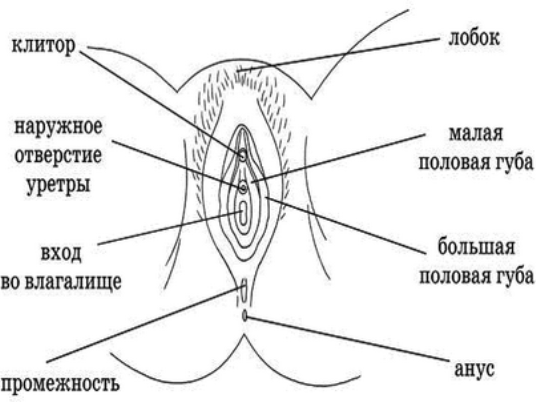
The pubis is the lowest part of the abdominal wall and represents a kind of elevation. It covers the lone articulation and performs a protective function, thanks to a large layer of fat. During puberty pubis is covered with hair.
The large labia are paired creases of the skin, which limit the sexual gap on each side. As a rule, they are pigmented, have a well-pronounced subcutaneous fat layer. Front, closing, form anterior adhesion, and from behind - anterior, which borders directly on the anus.
Small labia are also, in fact, nothing more than skin folds. They are located on the inside of the large lips and completely covered with them. In front small lips pass into the clitoris, and behind merge with the large labia.
Clitoris in its internal structure is an analog of the male penis, and consists of cavernous bodies that accumulate blood during sexual intercourse and increase it in size. The mucous membrane of the clitoris is rich in nerves, vessels, sweaty and, along with them, sebaceous glands, which produce smegma - lubricant.
The hymen is a thin mucous membrane that protects the internal organs and vagina. At the first sexual contact, a rupture of the spleen occurs (defloration), which is accompanied by a small discharge of blood. After this, the woman retains only the remains of the hymen in the form of the so-called papillae.
What are the structure and functions of internal female genital organs?
The vagina, in its shape, resembles a hollow tube through which the external and internal genital organs communicate. The average length is 7-9 cm. During intercourse and during childbirth, it can increase, due to the presence of a large number of folds that are straightened.
The main female genital organ is the uterus, it has a rather complex structure. In appearance it looks like a pear. It consists of 3 departments: body, neck and neck. The walls of the uterus have a well developed muscular layer, which allows it to easily increase in size during pregnancy.
Uterus, or fallopian tubes, are paired organs that depart directly from the body of the uterus.
Ovaries are paired glands, the main function of which is the synthesis of estrogens and progesterone. It is from their work that the overall condition of the reproductive system also often depends.
Thus, we can say that this structure of female genital organs is correct, but in the human anatomy deviations are often possible, which are due to both heredity and external factors on the body.