Problems with color perception can not always be diagnosed on time, which significantly degrades the quality of life. The test for color blindness can detect this genetic disease in a short time without special ophthalmologic manipulations. There are several varieties of this procedure.
What are the tests for color blindness and color perception?
Such types of incorrect perception of color are known:
- protoanopia (disturbances with color perception in the red spectrum);
- deuteranopia (disorders with color perception in the green spectrum).
In addition, there is absolute color blindness, in which people see the surrounding reality in black and white colors - monochromasia.
Normal perception of shades is called trichromasia.
The essence of the test for the examination of color blindness in an oculist consists in viewing a person with cards with images consisting of small colored circles. They form geometric figures and figures in such a way that people with normal color perception can see them, and patients with impairments either can not do this or observe other images.
Rubkin's test for color blindness
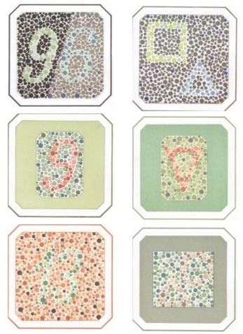
The study in question consists of viewing 23 cards. For each of them is allocated 9-10 seconds of time. It is important that the event is conducted in good light at rest. The image should be on the same level as the patient's eyes. Pictures should be viewed from left to right, from top to bottom.
On the first card - the number 69, on the second - a square and a triangle. They can be seen by people with normal color perception, and color-blind. These images are intended to illustrate the essence of the test to determine color blindness and identify the simulation.
Next, consider the cards in turn, the first number or figure visible to the trichromant:
- 3 - figure 9. With protoanopia and deuteronomy, the number 5 is visible.
- 4 - triangle. With blindness in the green and red part of the spectrum there is a circle.
- 5 - number 13. With deuteranopia and proto anopia - 6.
- 6 - a circle and a triangle. Color blinds do not see anything on the card.
- 7 - we can all see the number 9.
- 8 - number 5. It can be seen and people with color problems, but this is given with difficulty, it takes a long time to look closely.
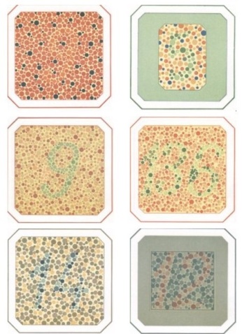
- 9 - figure 9 (visible to patients with deuteranopia). With protoanopia - 6 or 8.
- 10 is the number 136. Daltonics observe 66, 69 or 68.
- 11 - figure 14, visible to all.
- 12 - number 12, only deuteranopam and trichromantum are distinguishable. The Proto-Nopes can not see anything.
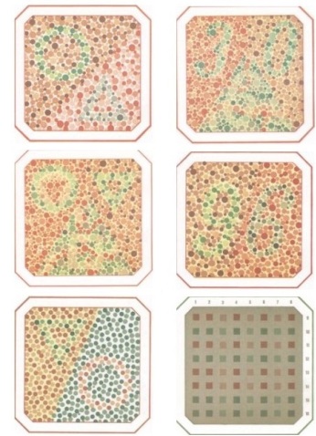
- 13 - triangle and circle. When violations with the perception of the green spectrum can be seen only the first figure, red - the second.
- 14 - in the upper part number 30. Proto-ops see there 10, and below - 6. Deuteranopes distinguish one at the top and 6 in the lower zone.
- 15 - a circle and a triangle (from above). Absence of perception of red shades allows to see two triangles at the top and a square from below, green ones - one triangle and a square.
- 16 - figure 96. With protoanopia, only nine are visible, deuteronopia - 6.
- 17 - a circle and a triangle. Changes in the visibility of the red part of the spectrum provide the visibility of the triangle, and the green one - only the circle.
- 18 - horizontal squares in one shade, vertical - multicolored. Deiteranopes see single-colored vertical rows 1, 2, 4, 6, 8, and all the horizontal ones seem to them to be multi-toned. Protanopes perceive 3, 5 and 7 vertical rows in one color, as well as horizontal lines of squares.
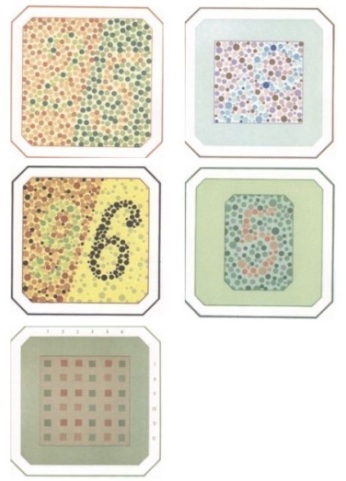
- 19 - the number 25. Color blinds see only 5.
- 20 - a circle and a triangle. Patients with impaired do not see anything.
- 21 is the figure 96, is also visible to proto-anopam. Deiteranopes do not see 9.
- 22 - number 5. It is difficult for the Daltons to discern the image or impossible.
- 23 - in the horizontal rows there are squares with different colors, and in vertical rows - the same shade. Patients with color blind perceive the card vice versa.
Rubkin's test for color blindness is sometimes called Rybkin's test (erroneously), it is important not to be confused with the tables of Ishihara or Ishihara. They look like Rubkin's cards, but instead of geometric figures, the Japanese ophthalmologist uses continuous curved lines.