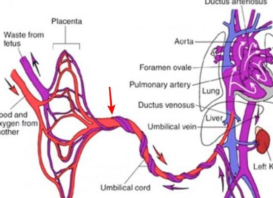
The supply of nutrients from the mother to the fetus, as well as the withdrawal of metabolic products is carried out with the help of the umbilical cord, which connects the placenta and the umbilical ring of the fetus.
Structure of the umbilical cord
It is important, from where the umbilical cord goes to the child: in norm it departs from the middle part of the placenta, although it is possible the marginal divergence - from some one of its edges, or the membrane attachment - the umbilical cord departs from the membranes from which the vessels from the placenta stretch. Its formation ends by 12 weeks, and the umbilical cord functions before the birth of the fetus. Usually the average length of the umbilical cord is from 40 to 70 cm, if less than 40 cm, it is a short umbilical cord , more than 70 cm is long.
How many vessels should an umbilical cord have?
Normally, the umbilical cord has three vessels: two arteries and a vein, between which there is a very strong substance, which prevents the vascular transmission in the umbilical cord: vartons jelly. But sometimes only 2 vessels are found in the umbilical cord, in 50% of cases it does not affect anything at all and the fetus develops normally. But, if the umbilical cord has only two vessels, it is necessary to examine the kidneys of the fetus, since this may be a sign of a congenital anomaly of the kidneys, or rather, a sign of the absence of one of the kidneys.
Node on the umbilical cord - what is it?
In the course of its development, the umbilical arteries grow and flex helically around the vein, and later the entire umbilical cord turns spirally. With the rapid growth of these vessels, the formation of coils from the vessels is possible, and with varicose veins of the umbilical vein, its node-like thickenings (false nodes of the umbilical cord). With false nodes, the blood flow in the umbilical cord is not impaired.
The true nodes of the umbilical cord are formed during fetal movements and during labor, but they rarely lead to negative consequences, only in the early stages of pregnancy, a tight knot may eventually cause atrophy of the varton jelly and cause a violation of the blood flow in the umbilical cord.
How dangerous is the cord with the umbilical cord?
During ultrasound investigation in the second half of pregnancy, usually the protocol records the presence of the umbilical cord near the neck. But, normally around the face of the child, there are often umbilical cords and it is necessary to check whether such a loop is around the neck. This is not always reliable in a routine study, but is clearly visible in Doppler. But the cord with the umbilical cord usually does not lead to negative consequences, if there are no other complications during childbirth, and is not a contraindication to natural delivery. But her presentation or prolapse of the umbilical cord loops from the birth canal is very dangerous for the fetus, since the compression of the umbilical cord between the birth canals and the fetus leads to asphyxia and fetal death in 90% of cases.